Sorry, but this retired science bod cannot think of a single good reason for thinking the Shroud image is on an impurity layer, whether it be starch, semi-degraded starch, soapwort, saponin, aloes, myrrh, whatever.
Nope. I'm not talking now about anecdotal evidence based on the supposition (at odds with the radiocarbon dating) that the Shroud is of 1st century AD provenance, when starch and/or natural soaps were allegedly used in spinning and weaving or bleaching.
I'm talking about hard chemical and other evidence, such as was acquired by the STURP team, to which the late Raymond N.Rogers was seconded as lead chemical investigator.
But you will not find Rogers' impurity coating thesis mentioned anywhere in the 1981 STURP summary of findings - and rightly so in my opinion.
OK, so playing hunches has a vital role in science ("framing hypotheses") but they have to be (a) testable in principle if they are to be taken seriously and (b) tested in practice before they can be accepted into the mainstream of scientific thinking.
Rogers' impurity coating idea not only lacks for hard experimental data, but the further development as his "Maillard reaction" hypothesis, proposing reaction between putrefaction amines(largely unspecified) and reducing sugars (largely unspecified) appears to this sceptic as pie-in-the-sky.
More importantly, the idea has never been properly put to the test, unless one accepts at face value the results of a questionable experiment involving ammonia and a contrived mix of (conveniently) partially-degraded starch ("dextrins"), providing the needed "reducing sugars" that intact starch is lacking. The yellow colour, assumed to be a Maillard product (evidence?) is open to alternative interpretation, given the absence of required controls, e.g. on pH (recalling that ammonia is a base, as well as a nitrogen donor). The elevated temperature (66 degrees Celsius) used to obtain a reasonable rate of reaction (or thermodynamic feasibility?) does nothing to cement credulity if intended as a model for natural processes occurring in and around a fairly recently-deceased shroud-wrapped cadaver.
Sorry, I'm going to have to end here for want of hard scientific data. Sorry if the title misled. Sometimes the means do justify the ends- like deliberately derailing a (now) driverless runaway locomotive before it's picked up too much speed.
Apologies in advance to those who think I have misjudged Raymond N.Rogers (RIP). Maybe I have overlooked crucial scientific evidence that underpins his "impurity layer" hypothesis. If so, please feel free to comment.
In the meantime, this Shroud investigator will continue to regard the hemicelluloses of the primary cell wall (PCW) as the most likely target for the image-forming mechanism, and sees no need, absolutely no need, to assume that untreated linen, free of starch and/or other processing aids, modern or classical, is incapable of accepting an image onto its intrinsic PCW, e.g. via direct physical contact with a heated metal template.
Postscript: I see that Adler's observation that diimide, NH=NH, bleaches the Shroud image, is being adduced as evidence in support of Rogers' ideas. I fail to see why (being unmoved by references to whether linen fibres look "clean" or "undamaged" before or after diimide treatment). What's the connection between events at the molecular level, and those at the gross one, with or without the benefit of a light microscope?
Second postscript: It's been suggested elsewhere that there is no way of telling whether the Shroud image is on the PCW or on a more superficial impurity coating. Admittedly that may be difficult using classical microscopy (atomic force microscopy is another matter). But it's not impossible using chemical means, though access to Shroud samples in the quantities required is as problematical as ever.
In fact I reminded folk last year of the technology that is available. It involves chemical hydrolysis of image v non-image areas via different graded acid-pretreatments, followed by gas-liquid or HPLC separation of constituent starch and cell wall sugars. An image on the PCW that resulted in partial or complete pyrolysis of the heat-sensitive hemicelluloses would be detected as a loss of hemicellulose markers (glucoxylans and other non-starch, non-cellulosic pentosan sugars etc) in the image-bearing samples relative to non-image controls. Alternatively, a putative image on a starch coating etc would be expected give the same profile of intact PCW sugars in image v non-image areas, with differences appearing in the additional complement of acquired starch etc, giving differences between the glucose recoveries.
About Me
- sciencebod
- Colin Berry, aka sciencebod, is a retired PhD researcher/teacher/academic who has worked in industry, medical schools, schools, food and biomedical research (mainly in the UK, but also in W.Africa and the United States). He's best known for his work on RESISTANT STARCH, recently described as "the trendiest form of dietary fibre".
See also his specialist Shroud of Turin blog on www.shroudofturinwithoutallthehype.wordpress.com
with over 350 postings to date. Oh, there's a second specialist site devoted to Stonehenge, Silbury Hill with some radical new thinking: https://sussingstonehenge.wordpress.com/
Popular Posts
-
 A note about the author ( added October 6, 2016 ): Thanks for visiting this post, penned some 6 years ago. It's still attracting 15-2...
 -
 New feature of this site (I can't speak for others): here's a LINK that takes you straight to Comments. Admittedly the c...
 -
 Update: added 10th April 2012 : My continuing research and ideas on the Turin Shroud are now to be found on two specialist sites, the more ...
 -
 Preamble (in red font ): break with previous practice: I have taken the tail end off the very long posting that preceded this one, and am ...
 -
 How could STURP's Final Conclusions have omitted to mention the most striking and unexpected feature of the Shroud's image - its...
 -
 Jokers(?) - left; victims - right So claims Helga Kotthoff, a German academic at the "Frieburg (sic) University if (sic) Educati...
 -
 Summary : This blogger’s hunch, indeed, growing conviction that Stonehenge, Avebury and other Neolithic sites were purpose-built for what i...
 -
 The basic principle - thermal imprinting from a 3D object (ed: things have moved on a bit since writing this post. For a more up-to-da...
 -
 Fig.1: Burning coal can produce flame, smoke AND smelly sulphurous fumes - even when well-supplied with oxygen. It's the result...
 -
 Note added October 7, 2016 : Dear reader. Please be aware that my 5 year Shroud investigation has come on considerably since...
Create one's own blog (age, class, gender no barrier)

It's really quite straightforward. All one has to do is to click on the photograph with that nice young man. One can then be part of the frightfully interesting Blogger community in just a couple of jiffs.

Acknowledgment

Image Courtesy, top row, from left: Reidar Hahn, Fermilab; BNL/STAR Collaboration; Kuhlman Laboratory, UNC; Fermilab; CERN; bottom row, from left: JHU; CERN; Fermilab; WRF; Shawn Rice, Purdue
What's the latest on the LHC?

LHC gets warning system upgrade : BBC 28 September 2009
Self-organization

From wiki entry on SELF ORGANIZATION: "As a result, processes considered part of thermodynamically open systems, such as biological processes that are constantly receiving, transforming and dissipating chemical energy (and even the earth itself which is constantly receiving and dissipating solar energy), can and do exhibit properties of self organization far from thermodynamic equilibrium."
How far away should your off-licence be for a bottle of wine to be energy-neutral?

Answer: about 3 miles, taken at standard walking pace (6 mile round trip)
What do these two have in common?
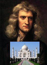
Answer: both arrived in this world about the same time. Sir Isaac Newton was born on 4th Jan 1643 (new style*). The Taj Mahal had a 20 year gestation period, centred on approximately the same year. Click on piccy for an older post .* Or Christmas Day, 1642, depending which dating system one uses.
Is interstellar space travel feasible?

The nearest star (more correctly, star system, since it's 3 stars, a binary and a smaller satellite star) is Alpha Centauri. The average distance from Earth is 4.3 light years. Suppose technology allows us one day to achieve an interstellar cruising speed of half the speed of light. A comfortable acceleration of g (simulating Earth's gravity) would take a year, with another year to slow down comfortably. The entire journey from Earth would take a minimum of 10 years approximately. Having arrived at one's destination, it would take 4.3 years to send a radio postcard (" Hello Mum and Dad. Have arrived safely, and am now looking for a habitable planet. Am hoping it's hiding behind Proxima. Have looked everywhere else... Would die for some Cheddar cheese... ")
What causes weather?

Could you answer that question in just 7 words, ie " weather is due to...? Need some help, " Weather is due to t- - u - - - - - - h - - - - - - o - t - - E- - - -'s s - - - - - - ." The National Curriculum (England and Wales) does have its uses, but there are many more such simple principles, expressed in a minimum of words, that could be usefully incorporated.
No comments:
Post a Comment